Introduction
Posterior Vitreous Detachment (PVD) is a common eye condition that occurs as a natural part of the aging process. It involves the separation of the gel-like substance called the vitreous from the retina, the light-sensitive layer at the back of the eye. This article aims to provide a comprehensive understanding of PVD, its causes, symptoms, treatment options, and potential complications. So, let's dive into the world of posterior vitreous detachment and unravel its mysteries.
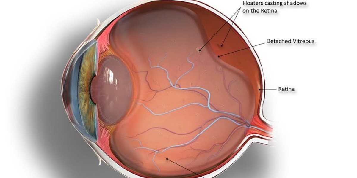
What Is Posterior Vitreous Detachment?
Posterior Vitreous Detachment, often abbreviated as PVD, refers to the separation of the vitreous gel from the retina. The vitreous humor, a transparent gel-like substance, fills the space between the lens and the retina in the eye. It plays a crucial role in maintaining the shape of the eye and transmitting light to the retina.
Symptoms of Posterior Vitreous Detachment
The onset of PVD can be characterized by various symptoms, which may vary from person to person. Some common symptoms include:
1. Floaters: Floaters are tiny specks or cobweb-like structures that appear to float in the field of vision. They are more noticeable when looking at a bright background, such as a clear sky or a white wall.
2. Flashes: Flashes of light are another common symptom of PVD. These flashes can occur spontaneously and are often described as brief and localized bursts of light.
3. Shadow or Curtain: In some cases, individuals may experience the perception of a shadow or curtain-like obstruction in their field of vision. This may be an indication of a more severe form of PVD and requires immediate medical attention.
4. Decreased Visual Acuity: PVD can lead to a temporary decrease in visual acuity, making it difficult to see objects clearly. This is often a result of floaters or other visual disturbances caused by the detachment of the vitreous gel.
Causes of Posterior Vitreous Detachment
Posterior Vitreous Detachment is primarily a natural aging process. As we grow older, the vitreous gel undergoes changes in its consistency and structure, making it more prone to detachment. However, certain factors may increase the risk of developing PVD. These include:
1. Age: PVD is more prevalent in individuals above the age of 50. The chances of experiencing PVD increase with age due to the natural changes occurring in the vitreous gel.
2. Myopia: High myopia, or nearsightedness, can contribute to an increased risk of developing PVD. The elongation of the eyeball associated with myopia can lead to a higher likelihood of vitreous detachment.
3. Eye Trauma: Significant eye trauma, such as a direct blow to the eye or an injury during eye surgery, can induce the separation of the vitreous gel from the retina, resulting in PVD.
4. Eye Surgery: Certain types of eye surgeries, such as cataract surgery, can predispose individuals to PVD. It is important to discuss the potential risks with an ophthalmologist before undergoing any surgical procedures.
Diagnosing Posterior Vitreous Detachment
If you experience any symptoms suggestive of PVD, it is crucial to seek prompt medical attention. An eye specialist, such as an ophthalmologist, can perform a comprehensive eye examination to diagnose the condition. The examination may involve the following:
1. Visual Acuity Test: This test measures the clarity of your vision using an eye chart. It helps determine if the detachment of the vitreous gel is affecting your visual acuity.
2. Dilated Eye Examination: In this examination, eye drops are used to dilate the pupil, allowing the ophthalmologist to examine the retina and the vitreous gel more closely. This can help confirm the presence of PVD and rule out any other underlying eye conditions.
3. Ultrasound Imaging: In some cases, ultrasound imaging may be used to obtain a more detailed view of the structures inside the eye. This can be particularly helpful if there are challenges in conducting a thorough examination using traditional methods.
Our Best Eye Surgeries in Delhi include:
Refractive Surgery
Cataract Surgery
Contact Lens
Laser Cataract Surgery
Retina Surgery
Glaucoma Treatment
Phacoemulsification Surgery
Smile Eye Surgery
Lasik Eye Surgery
Squint Treatment
Cornea Transplant
Pediatric Ophthalmology
Oculoplasty And Aesthetic
Visit our Website: Bharti Eye Foundation